Coming from Blindsight, we see that there are infact other parts of the brain that help process visuals.
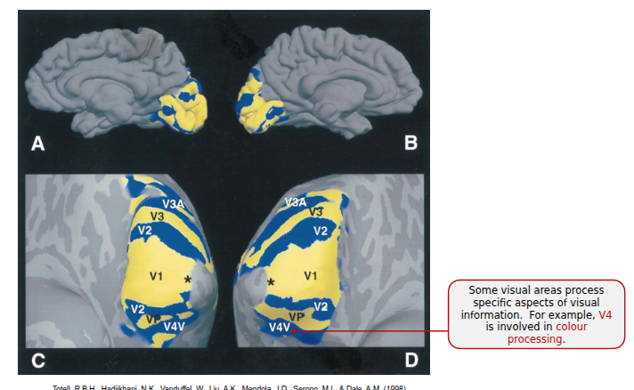
Primary and Secondary Visual Areas
Some visal areas process specific aspects of visual information.
- the V4 is involved in colour processing
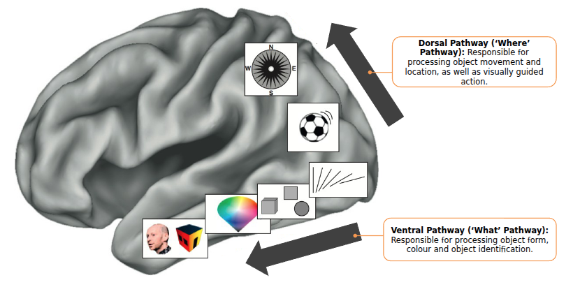
Ventral and Dorsal streams
- The ventral pathway is known as the ‘what pathway’, responsible for processing object’s form, color and identification
- The farther down we do in the ventral direction, the higher-order the pattern recognition is
- The dorsal pathway is known as the ‘where pathway’, responsible for locality of the object. Also helps with visually guided action like dodging a ball.
Ventral Temporal Cortex (bottom on the temporal Cortex)
temporalLobe The ventral temporal cortex has distinct specialized regions for different object categories.
- Fusiform Face Area responds specifically when processing faces and animate objects
- Parahippocampal Place Area responds specifically to places and inanimate objects
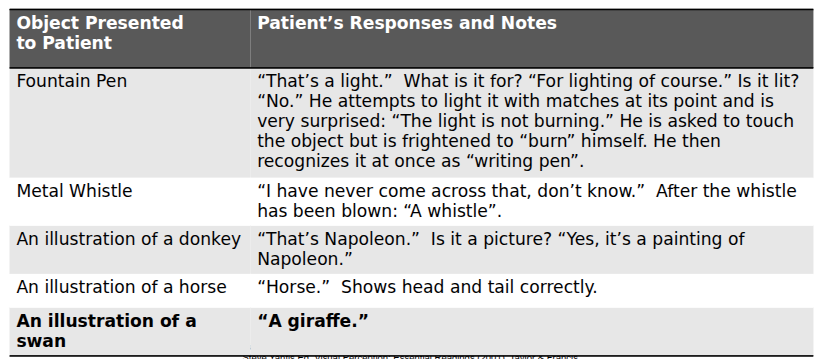
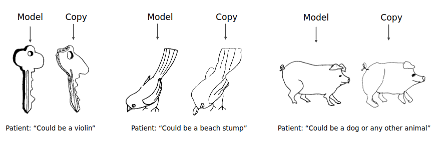
Visual Agnosia
An inability to recognize objects even though the patient can see them. Often involes damage to teh ventral temporal lobe.
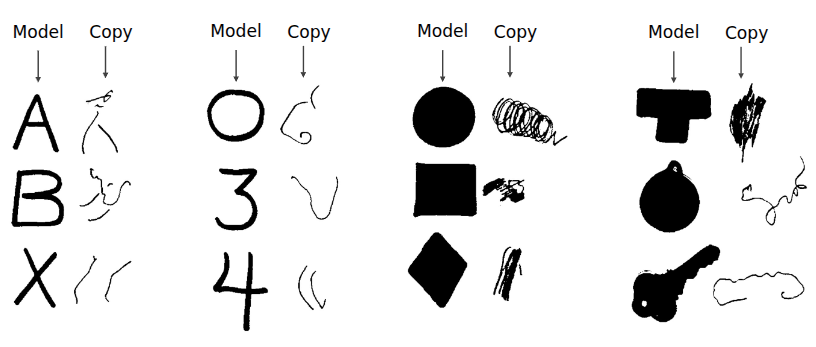
Apperceptive Agnosia
Inability to copy what they visualize.
Associative Agnosia
Inability to associate what they are visualizing to an object (even though they can copy it).
Patient D.F
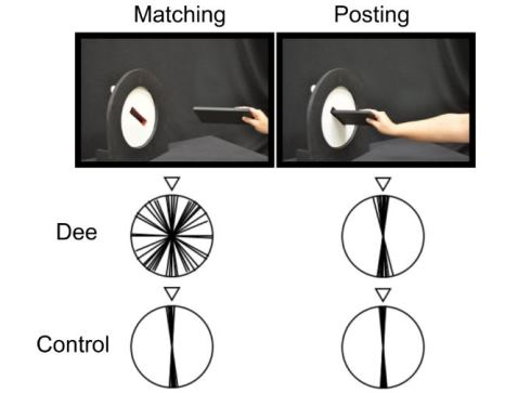 It consists of two tasks, one is to line up the slot with the right orientation as shown with the white tick. The other is to insert a card into the slot.
Patient DF was no able to align the slot properly (lacked the reasoning for shape and orientation BUT he was able to insert the post card in the slot really well (his actions based on orientation were still working).
Prosopagnosia
A type of visual agnosia where patients can see facial features but cannot attribute them to someone. They have to recognize people by their voice, gait, or clothing
- They lose their ability to see faces holistically
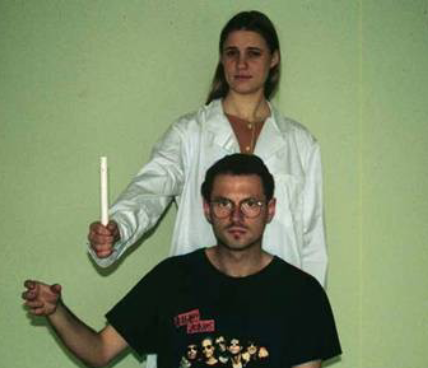
Optical Ataxia
Patients who have problems with visually guided reaching. Especially when the obejct is in their peripheral vision.
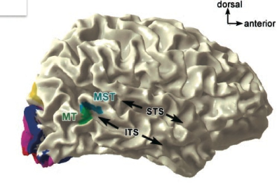
Motion
There are two distinct spots in the temporalLobe that deal with motion
Middle-temporal Cortex (MT/V5)
Has a retinotopic organization (there is a direct map between visual field and neuron topology).
Responds to direction of movement, specific speed, change in speed, object movement relative of the background
Medial Superior Temporal Cortex (MST)
Coarse retinotopic map (there is a coarse mapping between locations in the visualfield and the neuron topology). These neurons have a larger receptive field as a result.
Responds to:
- Scene expansion and contraction (Dorsal Part)
- Object movement relative to the background (Lateral-ventral part)
Visual Motion Blindness (Akinetopsia)
Patients lack the ability to perceive motion.
- cannot understand things moving, they just seem to teleport.
- they see freeze frames
- And their consciousness actually views the world as freeze frames, it doesn’t have the ability to stitch a motion-rich world for them to perceive.